High and Low Pressure Headache
Idiopathic Intracranial Hypertension (IIH)
- Headache worsens on awakening
- Pulsatile tinnitus
- Papilledema
Low Pressure Headache
Occur on rising and/or later in the day
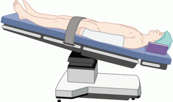
Headache triggered by standing. In clinic, put patient in Trendelenburg (the body is laid supine, or flat on the back with the feet higher than the head by 15-30 degrees) 10 min, headache should resolve. Intracranial Spinal CSF leak, spontaneous or trauma (may be trivial) or valsalva. Ehlers-Danlos or joint laxity. Fragile dura. Brain MRI with contrast: dural thickening, brain sag. Do not confuse with Chiari malformation. Don't do LP routinely; if you do, use small needle, 24 gauge. Low pressure not always present: <60 mm. Cervical cord or thoracic cord. Identification of leak: CT and MR myelogram (intrathecal gadolinium, off label use).
Headache triggered by standing. In clinic, put patient in Trendelenburg (the body is laid supine, or flat on the back with the feet higher than the head by 15-30 degrees) 10 min, headache should resolve. Intracranial Spinal CSF leak, spontaneous or trauma (may be trivial) or valsalva. Ehlers-Danlos or joint laxity. Fragile dura. Brain MRI with contrast: dural thickening, brain sag. Do not confuse with Chiari malformation. Don't do LP routinely; if you do, use small needle, 24 gauge. Low pressure not always present: <60 mm. Cervical cord or thoracic cord. Identification of leak: CT and MR myelogram (intrathecal gadolinium, off label use).
Leak in the Spinal Column, Most Often Low Cervical or Thoracic
Website for Patients
https://spinalcsfleak.org/
Key Factors
Bilateral more common than unilateral.
Risk factors: joint hypermobility, previous lumbar puncture, epidural or spinal anesthesia, known disc disease, or a personal or family history of retinal detachment at a young age, aneurysm, dissection, or valvular heart disease.
Physical Examination
Conservative measures don’t work very well. Even if a leak site hasn’t been identified, treat with a high-volume epidural CT-guided targeted blood patch with fibrin sealant. (Relief about a third of the time each time you do it)
Website for Patients
https://spinalcsfleak.org/
Key Factors
- Postural, end-of-the-day, and Valsalva components to the headache are present
- Joint hypermobility
- Orthostatic or gets worse at end of day. (Longer patient has SIH, the less likely there is a postural component.)
- Majority of patients are awakened by headache in middle of night.
- Headache is often exertional and worsens with Valsalva including coughing, sneezing, lifting, bending forward, straining, singing, or sexual activity.
- Caffeine often works very well.
- May be thunderclap in onset but not necessarily.
- Tinnitus
- Abnormal hearing as if underwater
- Neck pain, imbalance
- Pain between shoulder blades
- Blurred or double vision.
Bilateral more common than unilateral.
Risk factors: joint hypermobility, previous lumbar puncture, epidural or spinal anesthesia, known disc disease, or a personal or family history of retinal detachment at a young age, aneurysm, dissection, or valvular heart disease.
Physical Examination
- Joint hypermobility
- Spontaneous retinal venous pulsations indicative of normal CSF pressure are present in the eyes
- Put patient in 5 degrees of Trendelenburg position for 5-10 minutes to see if that improves the headache and other symptoms.
- Brain MRI with gadolinium enhancement (normal in 30% of affected patients)
- No consensus when the brain MRI is negative. Can do (1) CT with or without MR myelography, or (2) T2-weighted spine MRI. (No leak is found in about half of individuals with SIH.)
Conservative measures don’t work very well. Even if a leak site hasn’t been identified, treat with a high-volume epidural CT-guided targeted blood patch with fibrin sealant. (Relief about a third of the time each time you do it)
CSF Leak Symptoms by Frequency
HEADACHE AND PAIN
HEADACHE AND PAIN
- Orthostatic headache – 92%
- Neck / interscapular pain – 33%
- Daily headache
- 2nd half of the day headache
- Exertional / valslva headache
- Paradoxical orthostatic
- Nausea – 51%
- Hearing/tinnitus/ear symptoms – 33%
- Dizziness, vertigo – 18%
- Visual symptoms
- Altered consciousness
- Extrapyramidal
- Cognitive (frontotemporal)
